Interventional Radiology
About Interventional Radiology
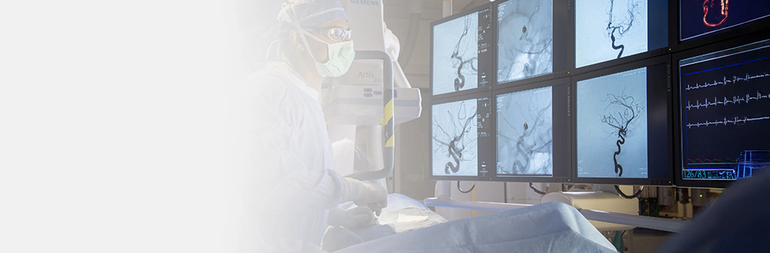
Our Interventional Radiology Department provides minimally invasive, image guided procedures for accurate diagnosis and targeted treatment. Using advanced imaging technologies, our specialists deliver safe and precise care with shorter recovery times and reduced risks.
At University Hospital Sharjah, we offer a wide range of minimally invasive procedures supported by advanced imaging such as X ray fluoroscopy, ultrasound, CT, and MRI. These technologies allow our specialists to guide treatments with high accuracy while minimizing the need for traditional surgery.
Whether you are beginning image guided treatment or looking for less invasive options to manage complex conditions, our interventional radiology team provides tailored service care focused on safety, comfort, and better clinical outcomes.
Symptoms
You may benefit from minimally invasive, image-guided treatment with an interventional radiologist in Sharjah if you experience:
Visible Varicose Veins or Leg Swelling
These symptoms may indicate venous insufficiency or deep vein thrombosis, which can be treated with targeted, non-surgical techniques.
Chronic Pelvic Pain or Heavy Menstrual Bleeding
These may be signs of uterine fibroids or pelvic congestion syndrome—both manageable with minimally invasive procedures such as uterine fibroid embolization.
Suspected or Diagnosed Tumors
Interventional radiology offers image-guided biopsies, tumor ablation, or chemoembolization as part of cancer diagnosis and treatment.
Recurrent Urinary Tract Blockages or Kidney Stones
When stones or strictures interfere with kidney drainage, interventional techniques like nephrostomy tubes or stenting may be necessary.
Unexplained Gastrointestinal Distress
Conditions such as gastrointestinal bleeding, bowel obstruction, hemorrhoids, or liver disease may benefit from interventional imaging and procedures.
Peripheral Artery Disease
All types of endovascular procedures to treat stenosed or occluded arteries are available at our department.
Chronic Joint and Tendon Pain
We treat chronic musculoskeletal pain such as low back pain caused by degenerative disc disorders or joint pain related to osteoarthritis.
Benign Prostate Hypertrophy and Varicocele
Minimally invasive, image-guided techniques offer effective treatment without traditional surgery.
Stroke Symptoms or Neurovascular Complications
Emergent or preventive care for vascular blockages may involve advanced, image-guided catheter-based treatments.
What Might Be Causing It
Many conditions managed by interventional radiologists arise from underlying or chronic health issues, including:
Specialized Interventional Radiology Department Services & Procedures
Our Interventional Radiology Department at UHS delivers precise, image-guided care for a wide range of conditions using advanced tools and techniques. These include:
Why Choose University Hospital Sharjah for Interventional Radiology?
Our board-certified interventional radiologists in Sharjah bring advanced expertise in minimally invasive procedures supported by modern imaging technologies. Using precise, image-guided techniques, our team performs treatments such as tumor ablation, biopsies, and angioplasty safely and effectively.
We offer gentler alternatives to open surgery, including uterine fibroid and varicocele embolization, with less pain and faster recovery. Through close collaboration with gynecology, oncology, neurology, and vascular surgery, every patient receives coordinated, comprehensive care. Contact us today to book your consultation at University Hospital Sharjah.